2024
|
|
|
|
 |
 |
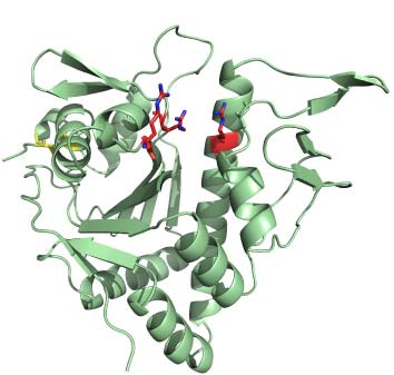
Molecular basis of hemoglobin capture by
Corynebacterium diphtheriae
|
|
|
|
 |
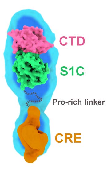
Insight into the autoproteolysis mechanism of the RsgI9 anti-σ factor
from Clostridium thermocellum (ref. 124)
|
|
|
|
2023
|
|
|
|
|
 |
The Shr receptor from Streptococcus pyogenes uses a 'cap and release'
mechanism to acquire heme-iron from human hemoglobin (ref. 120)
|
|
|
|
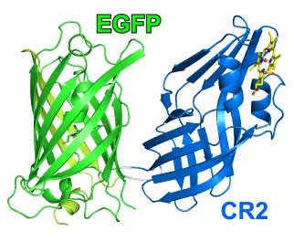 |
Development and atomic structure of a new fluorescence-based sensor
to probe heme transfer in bacterial pathogens (ref. 123)
|
|
|
|
 |
The basal and major pilins in the Corynebacterium diphtheriae SpaA pilus
adopt similar structures that competitively react with the pilin polymerase
(ref. 121)
|
|
|
|
2022
|
|
|
|
|
 |
Insight into the molecular basis of substrate recognition by the
wall teichoic acid glycosyltransferase TagA (ref. 117)
|
|
|
|
 |
The structure of the Clostridium thermocellum RsgI9 ectodomain
provides insight into the mechanism of biomass sensing (ref. 118)
|
|
|
|
2021
|
|
|
|
|
 |
Sortase-assembled pili in Corynebacterium diphtheriae are built using
a latch mechanism (ref. 115)
|
|
|
|
2019
|
|
|
|
|
 |
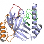
Structure and mechanism of LcpA, a phosphotransferase that mediates
glycosylation of a Gram-positive bacterial cell wall-anchored protein
(ref. 105)
|
|
|
|
2018
|
|
|
|
|
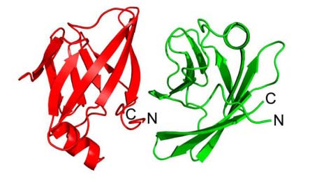 |
The Streptococcus pyogenes Shr protein captures human hemoglobin
using two structurally unique binding domains (ref.104)
|
|
|
|
2017
|
|
|
|
|

|
NMR structure-based optimization of pyridazinone class
Staphylococcus aureus Sortase A inhibitors (ref. 93)
|
|
|
|
2016
|
|
|
|
|
 |
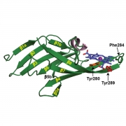
Crystal Structure of the Streptomyces coelicolor Sortase E1
Transpeptidase Provides Insight into the Binding Mode of
the Novel Class E Sorting Signal (ref. 92)
|
|
|
-
|
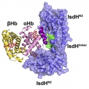
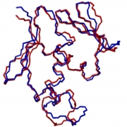
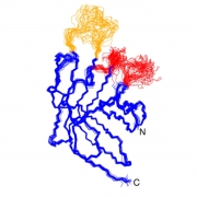
The PRE-Derived NMR Model of the 38.8-kDa Tri-Domain IsdH
Protein from Staphylococcus aureus Suggests That It Adaptively
Recognizes Human Hemoglobin. (ref. 83)
|
|
|
2015
|
|
|
|
|
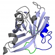
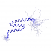
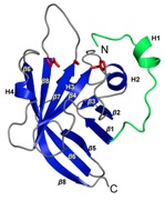
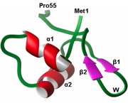
Solution structure of the PhoP DNA-binding domain from
Mycobacterium tuberculosis. (ref. 86)
|
|
|
2014
|
|
|
|
 |
Novel mechanism of hemin capture by Hbp2, the hemoglobin-binding
hemophore from Listeria monocytogenes. (ref. 82)
|
|
|
 |
Structural and computational studies of the Staphylococcus aureus
sortase B-substrate complex reveal a substrate-stabilized oxyanion hole.
(ref. 79)
|
|
|
2013
|
|
|
|
 |
Staphylococcus aureus uses a novel multidomain receptor to break
apart human hemoglobin and steal its heme. (ref. 74)
|
|
|
2012
|
|
|
|
 |
Solution structure of the sortase required for efficient production
of infectious Bacillus anthracis spores. (ref. 88)
|
|
|
2011
|
|
|
|
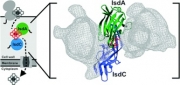 |
Transient weak protein-protein complexes transfer heme across
the cell wall of Staphylococcus aureus. (ref. 68)
|
|
|
|
Solution structure of the sortase enzyme from Bacillus anthracis
contains a novel active site architecture (ref. 62)
|
|
|

|
Structure of the sortase-substrate complex from
Staphylococcus aureus (ref. 59)
|
|
|
|
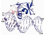
NMR structure of the amino-terminal domain of the
lambda integrase protein in complex with DNA (ref. 60)
|
|
|
2008
|
|
|
|

|
Structure of the IsdC-ZnPPIX complex (ref. 56)
|
|
|
2007
|
|
|
|
|
Crystal structure of the cooperative Xis-DNA complex (ref. 51)
|
|
|
|
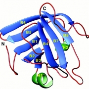
NMR structure of the ORF1 RNA binding domain from LINE-1 (ref. 54)
|
|
|
2006
|
|
|
|
|
NMR structure of the IsdH hemoglobin receptor (ref. 49)
|
|
|
2005
|
|
|
|
|
NMR structure of the Tn916 Xis protein (ref. 44)
|
|
|
|
Crystal structure of single Xis-DNA complex (ref. 41)
|
|
|
2002
|
|
|
|
|
NMR structure of the ARID-DNA complex (ref. 31)
|
|
|

|
NMR structure of the lambda integrase DNA binding domain (ref. 32)
|
|
|
|
NMR structure of the lambda Xis protein (ref. 34)
|
|
|
2001
|
|
|
|
|
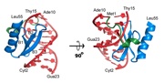
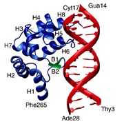
NMR structure of the Mu Repressor-DNA complex (ref. 29)
|
|
|
|
NMR structure of Sortase (ref. 26)
|
|
|
|
|
|